Dive into the fascinating world of cell division with onion root tip 1000x labeled! This guide unravels the intricate processes of mitosis, providing a comprehensive exploration of this fundamental biological phenomenon.
Using onion root tips as a model system, we’ll embark on a journey to understand the stages of mitosis, the role of the spindle apparatus and chromosomes, and the significance of these studies in various fields.
Introduction to Onion Root Tip
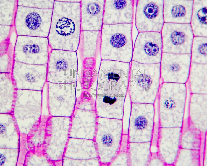
Onion root tips have emerged as an invaluable tool for studying mitosis, the fundamental process of cell division. Their significance stems from their inherent characteristics, which render them particularly suitable for cytological investigations.
The advantages of utilizing onion root tips for cytological studies are multifaceted. Firstly, onion root tips exhibit a high rate of cell division, providing an ample supply of cells undergoing mitosis. Secondly, the cells within onion root tips are relatively large, allowing for clear visualization of chromosomal structures during cell division.
Moreover, the root tips are easily accessible and can be readily collected for experimentation.
Advantages of Onion Root Tips for Cytological Studies
- High rate of cell division ensures a plentiful supply of cells undergoing mitosis.
- Large cell size facilitates clear visualization of chromosomal structures during cell division.
- Easily accessible and readily collected for experimentation.
Preparation of Onion Root Tip Slides
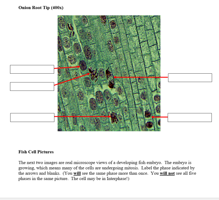
To study the detailed structure of onion root tip cells, we need to prepare slides of the root tips. Here’s a step-by-step guide to prepare onion root tip slides for microscopy:
Collection of Root Tips
1. Collect actively growing onion roots from a healthy onion bulb.
2. Cut off approximately 1-2 cm of the root tips using a sharp razor blade or scalpel.
Fixation of Root Tips
3. Place the root tips in a fixative solution, such as Carnoy’s solution (3:1 ethanol:acetic acid), for 24-48 hours. This step preserves the cell structure.
Staining of Root Tips
4. After fixation, rinse the root tips with distilled water.
5. Stain the root tips with a suitable stain, such as acetocarmine or Feulgen stain. Acetocarmine stains the chromosomes, while Feulgen stain specifically stains DNA.
6. Rinse the root tips with distilled water again.
Squashing of Root Tips
7. Place a stained root tip on a microscope slide and add a drop of acetocarmine or Feulgen stain.
In a study of onion root tip 1000x labeled, researchers were able to identify various cell structures. If you’re looking for additional resources on this topic, check out the vhl spanish 1 answer key . Continuing our exploration of the onion root tip 1000x labeled, we can observe the nucleus, vacuoles, and chromosomes.
8. Gently tap the root tip with a coverslip to spread the cells.
9. Blot away excess stain using a tissue paper.
10. Observe the slide under a microscope using the appropriate magnification.
Observation and Analysis of Onion Root Tip Cells
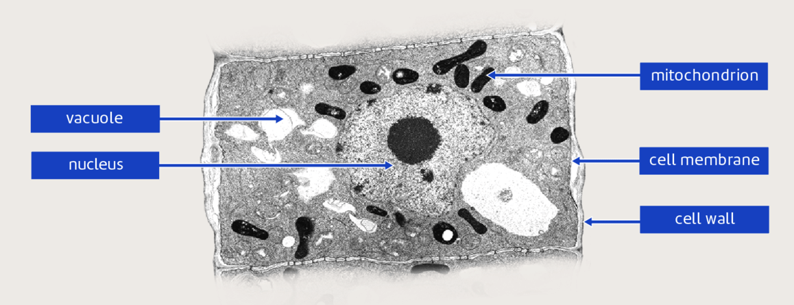
Observing and analyzing onion root tip cells under a microscope is a fundamental technique in cell biology. It allows researchers to study the structure and function of plant cells, including their chromosomes and the process of cell division.
Onion root tips are an excellent material for studying mitosis because they are readily available, easy to prepare, and have a high mitotic index. In this guide, we will provide a comprehensive overview of the steps involved in observing and analyzing onion root tip cells under a microscope, including the identification of different stages of mitosis.
Identification of Mitosis Stages
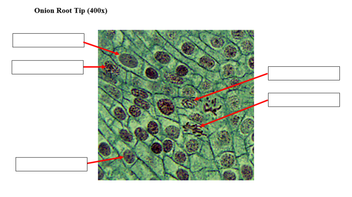
Mitosis is the process by which a cell divides into two identical daughter cells. It consists of four distinct stages: prophase, metaphase, anaphase, and telophase. Each stage has its own unique characteristics, which can be identified under a microscope.
| Stage | Key Characteristics |
|---|---|
| Prophase | Chromosomes become visible and condense. Nuclear envelope breaks down. Spindle fibers form. |
| Metaphase | Chromosomes align at the equator of the cell. Spindle fibers attach to the chromosomes. |
| Anaphase | Sister chromatids separate and move to opposite poles of the cell. |
| Telophase | Chromosomes reach the poles of the cell. Nuclear envelope reforms. Spindle fibers disappear. |
By carefully observing and analyzing onion root tip cells under a microscope, researchers can gain valuable insights into the process of cell division and the structure and function of plant cells.
Mitosis in Onion Root Tip Cells
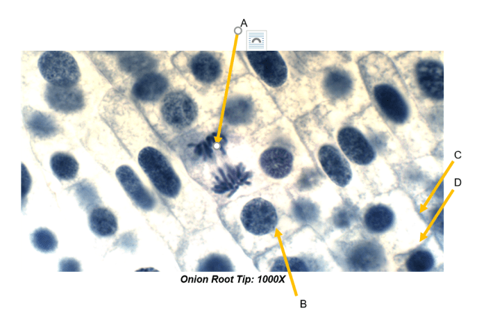
Mitosis is the process of cell division that results in two genetically identical daughter cells. It is a continuous process, but for the sake of study, it can be divided into four distinct stages: prophase, metaphase, anaphase, and telophase.
During prophase, the chromatin condenses into visible chromosomes, and the nuclear envelope breaks down. The spindle apparatus, a complex structure made of microtubules, begins to form. The spindle apparatus attaches to the chromosomes at their centromeres.
Metaphase
In metaphase, the chromosomes line up in the center of the cell. The spindle apparatus is fully formed and attached to the chromosomes.
Anaphase
In anaphase, the sister chromatids of each chromosome separate and move to opposite poles of the cell. The spindle apparatus shortens, pulling the chromosomes apart.
Telophase
In telophase, the chromosomes reach the poles of the cell and begin to decondense. The nuclear envelope reforms around each set of chromosomes. The spindle apparatus breaks down. Cytokinesis, the division of the cytoplasm, then occurs, resulting in two genetically identical daughter cells.
Mitosis is an essential process for growth, development, and repair. It is also the process by which new cells are produced to replace old or damaged cells.
Applications of Onion Root Tip Studies: Onion Root Tip 1000x Labeled
Onion root tip studies have proven to be invaluable in various scientific disciplines, particularly in the fields of cell biology, genetics, and education. These studies have significantly contributed to our understanding of cell division and the fundamental principles of genetics.
Applications in Cell Biology
Onion root tip studies have been extensively used to investigate the process of cell division, known as mitosis. The root tips provide an ideal model for studying mitosis due to their rapid cell division and the clear visibility of chromosomes during different stages of the process.
Researchers have utilized onion root tips to observe and analyze the intricate events of mitosis, including chromosome condensation, spindle fiber formation, and cytokinesis. These studies have provided crucial insights into the mechanisms and regulation of cell division, which are essential for growth, development, and tissue repair.
Applications in Genetics, Onion root tip 1000x labeled
Onion root tip studies have also played a significant role in advancing our understanding of genetics. By analyzing the chromosomes in onion root tip cells, researchers have been able to identify and map genes, study genetic mutations, and investigate the inheritance patterns of traits.
These studies have contributed to the development of genetic maps, which are essential for understanding the organization and function of genes within an organism’s genome.
Applications in Education
Onion root tip studies have become a cornerstone of biology education at various levels. They provide a hands-on and engaging way for students to learn about cell division, genetics, and microscopy techniques. Students can prepare and observe onion root tip slides to visualize different stages of mitosis and identify chromosomes.
These studies help students develop a deeper understanding of the fundamental processes of life and prepare them for further studies in biology and related fields.
FAQ Section
What are the advantages of using onion root tips for studying mitosis?
Onion root tips are readily available, easy to collect, and have a high mitotic index, making them ideal for studying cell division.
How can I prepare onion root tip slides for microscopy?
To prepare onion root tip slides, collect root tips, fix them in a preservative, and stain them with a dye to visualize the chromosomes.
What are the key stages of mitosis?
Mitosis consists of prophase, metaphase, anaphase, and telophase, each with distinct chromosomal arrangements and spindle fiber dynamics.